TCL1 Monoclonal Antibody (eBio1-21 (1-21)), PE-Cyanine7, eBioscience
PRODUCT DETAILS
Host: Mouse
Isotype: IgG2b, kappa
Clonality: Monoclonal
Clone: eBio1-21 (1-21)
Format: PE-Cyanine7
Reactivity: Hu
Application: Flow Cytometry
Tested Dilution: 5 µL (0.25 µg)/test
Concentration: 5 μL/Test
Storage: 4°C, store in dark, DO NOT FREEZE!
Formulation: PBS with BSA and 0.09% sodium azide; pH 7.2
Purification: Affinity chromatography
Data Sheet: TDS
Specific Information
Description: The eBio1-21 antibody reacts with human T cell leukemia/lymphoma 1 (TCL1), a 14 kDa proto-oncogene product with a suggested role in intracellular regulation of T cell signalling. TCL1 was identified as the oncogene located at the 14q32.1 chromosome breakpoint region in T-cell prolymphocytic leukemia (T-PLL). In T-PLL, TCL1 is overexpressed as a result of an inversion or a reciprocal translocation, by juxtaposition to the T-cell receptor promoter/enhancer elements. TCL1 binds to the pleckstrin homology domain of Akt (protein kinase B) family proteins, which facilitates Akt dimerization and activity. By increasing Akt activity TCL1 may enhance the serine/threonine phosphorylation of major Akt signaling substrates, such as Ikk complex, mTOR, BAD, p70S6 kinase, FOXO transcription factors, and GSK3beta. These substrates regulate cellular differentiation, growth, survival, and metabolism.
Besides its tumorigenic role in T-PLL, TCL1 is normally expressed in the CD3-CD4-CD8- subset of thymic precursors in the T cell lineage, the plasmacytoid subset of dendritic cells, stimulated (not resting) mature T cells, and B cells up to the germinal center stage of maturation. TCL1 is inappropriately expressed by chromosome rearrangements that lead to pre-malignant clonal T cell expansions and mature T cell tumors. TCL1 shows a regulated expression pattern in chronic lymphocytic leukemia (CLL).
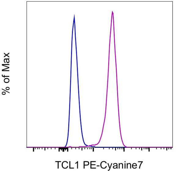
Applications Reported: This eBio1-21 antibody has been reported for use in intracellular staining followed by flow cytometric analysis.
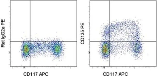
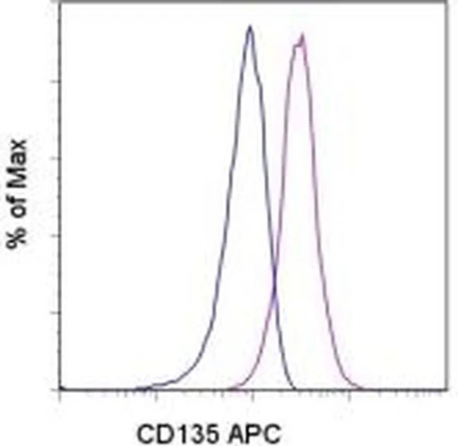
Applications Tested: This eBio1-21 antibody has been pre-diluted and tested by intracellular staining followed by flow cytometric analysis of Daudi cells using the Intracellular Fixation & Permeabilization Buffer Set (Product # 88-8824-00) and protocol. Please refer to "Staining Intracellular Antigens for Flow Cytometry, Protocol A: Two step protocol for intracellular (cytoplasmic) proteins" located at www.thermofisher.com/flowprotocols . This may be used at 5 µL (0.25 µg) per test. A test is defined as the amount (µg) of antibody that will stain a cell sample in a final volume of 100 µL. Cell number should be determined empirically but can range from 10^5 to 10^8 cells/test.
Light sensitivity: This tandem dye is sensitive to photo-induced oxidation. Please protect this vial and stained samples from light.
Fixation: Samples can be stored in IC Fixation Buffer (Product # 00-8222-49) (100 µL of cell sample + 100 µL of IC Fixation Buffer) or 1-step Fix/Lyse Solution (Product # 00-5333-57) for up to 3 days in the dark at 4°C with minimal impact on brightness and FRET efficiency/compensation. Some generalizations regarding fluorophore performance after fixation can be made, but clone specific performance should be determined empirically.
Excitation: 488-561 nm; Emission: 775 nm; Laser: Blue Laser, Green Laser, Yellow-Green Laser.
For Research Use Only. Not for use in diagnostic procedures. Not for resale without express authorization.
PRODUCT DETAILS
Host: Mouse
Isotype: IgG2b, kappa
Clonality: Monoclonal
Clone: eBio1-21 (1-21)
Format: PE-Cyanine7
Reactivity: Hu
Application: Flow Cytometry
Tested Dilution: 5 µL (0.25 µg)/test
Concentration: 5 μL/Test
Storage: 4°C, store in dark, DO NOT FREEZE!
Formulation: PBS with BSA and 0.09% sodium azide; pH 7.2
Purification: Affinity chromatography
Data Sheet: TDS
Specific Information
Description: The eBio1-21 antibody reacts with human T cell leukemia/lymphoma 1 (TCL1), a 14 kDa proto-oncogene product with a suggested role in intracellular regulation of T cell signalling. TCL1 was identified as the oncogene located at the 14q32.1 chromosome breakpoint region in T-cell prolymphocytic leukemia (T-PLL). In T-PLL, TCL1 is overexpressed as a result of an inversion or a reciprocal translocation, by juxtaposition to the T-cell receptor promoter/enhancer elements. TCL1 binds to the pleckstrin homology domain of Akt (protein kinase B) family proteins, which facilitates Akt dimerization and activity. By increasing Akt activity TCL1 may enhance the serine/threonine phosphorylation of major Akt signaling substrates, such as Ikk complex, mTOR, BAD, p70S6 kinase, FOXO transcription factors, and GSK3beta. These substrates regulate cellular differentiation, growth, survival, and metabolism.
Besides its tumorigenic role in T-PLL, TCL1 is normally expressed in the CD3-CD4-CD8- subset of thymic precursors in the T cell lineage, the plasmacytoid subset of dendritic cells, stimulated (not resting) mature T cells, and B cells up to the germinal center stage of maturation. TCL1 is inappropriately expressed by chromosome rearrangements that lead to pre-malignant clonal T cell expansions and mature T cell tumors. TCL1 shows a regulated expression pattern in chronic lymphocytic leukemia (CLL).
Applications Reported: This eBio1-21 antibody has been reported for use in intracellular staining followed by flow cytometric analysis.
Applications Tested: This eBio1-21 antibody has been pre-diluted and tested by intracellular staining followed by flow cytometric analysis of Daudi cells using the Intracellular Fixation & Permeabilization Buffer Set (Product # 88-8824-00) and protocol. Please refer to "Staining Intracellular Antigens for Flow Cytometry, Protocol A: Two step protocol for intracellular (cytoplasmic) proteins" located at www.thermofisher.com/flowprotocols . This may be used at 5 µL (0.25 µg) per test. A test is defined as the amount (µg) of antibody that will stain a cell sample in a final volume of 100 µL. Cell number should be determined empirically but can range from 10^5 to 10^8 cells/test.
Light sensitivity: This tandem dye is sensitive to photo-induced oxidation. Please protect this vial and stained samples from light.
Fixation: Samples can be stored in IC Fixation Buffer (Product # 00-8222-49) (100 µL of cell sample + 100 µL of IC Fixation Buffer) or 1-step Fix/Lyse Solution (Product # 00-5333-57) for up to 3 days in the dark at 4°C with minimal impact on brightness and FRET efficiency/compensation. Some generalizations regarding fluorophore performance after fixation can be made, but clone specific performance should be determined empirically.
Excitation: 488-561 nm; Emission: 775 nm; Laser: Blue Laser, Green Laser, Yellow-Green Laser.
For Research Use Only. Not for use in diagnostic procedures. Not for resale without express authorization.
Original: $453.00
-70%$453.00
$135.90Description
PRODUCT DETAILS
Host: Mouse
Isotype: IgG2b, kappa
Clonality: Monoclonal
Clone: eBio1-21 (1-21)
Format: PE-Cyanine7
Reactivity: Hu
Application: Flow Cytometry
Tested Dilution: 5 µL (0.25 µg)/test
Concentration: 5 μL/Test
Storage: 4°C, store in dark, DO NOT FREEZE!
Formulation: PBS with BSA and 0.09% sodium azide; pH 7.2
Purification: Affinity chromatography
Data Sheet: TDS
Specific Information
Description: The eBio1-21 antibody reacts with human T cell leukemia/lymphoma 1 (TCL1), a 14 kDa proto-oncogene product with a suggested role in intracellular regulation of T cell signalling. TCL1 was identified as the oncogene located at the 14q32.1 chromosome breakpoint region in T-cell prolymphocytic leukemia (T-PLL). In T-PLL, TCL1 is overexpressed as a result of an inversion or a reciprocal translocation, by juxtaposition to the T-cell receptor promoter/enhancer elements. TCL1 binds to the pleckstrin homology domain of Akt (protein kinase B) family proteins, which facilitates Akt dimerization and activity. By increasing Akt activity TCL1 may enhance the serine/threonine phosphorylation of major Akt signaling substrates, such as Ikk complex, mTOR, BAD, p70S6 kinase, FOXO transcription factors, and GSK3beta. These substrates regulate cellular differentiation, growth, survival, and metabolism.
Besides its tumorigenic role in T-PLL, TCL1 is normally expressed in the CD3-CD4-CD8- subset of thymic precursors in the T cell lineage, the plasmacytoid subset of dendritic cells, stimulated (not resting) mature T cells, and B cells up to the germinal center stage of maturation. TCL1 is inappropriately expressed by chromosome rearrangements that lead to pre-malignant clonal T cell expansions and mature T cell tumors. TCL1 shows a regulated expression pattern in chronic lymphocytic leukemia (CLL).
Applications Reported: This eBio1-21 antibody has been reported for use in intracellular staining followed by flow cytometric analysis.
Applications Tested: This eBio1-21 antibody has been pre-diluted and tested by intracellular staining followed by flow cytometric analysis of Daudi cells using the Intracellular Fixation & Permeabilization Buffer Set (Product # 88-8824-00) and protocol. Please refer to "Staining Intracellular Antigens for Flow Cytometry, Protocol A: Two step protocol for intracellular (cytoplasmic) proteins" located at www.thermofisher.com/flowprotocols . This may be used at 5 µL (0.25 µg) per test. A test is defined as the amount (µg) of antibody that will stain a cell sample in a final volume of 100 µL. Cell number should be determined empirically but can range from 10^5 to 10^8 cells/test.
Light sensitivity: This tandem dye is sensitive to photo-induced oxidation. Please protect this vial and stained samples from light.
Fixation: Samples can be stored in IC Fixation Buffer (Product # 00-8222-49) (100 µL of cell sample + 100 µL of IC Fixation Buffer) or 1-step Fix/Lyse Solution (Product # 00-5333-57) for up to 3 days in the dark at 4°C with minimal impact on brightness and FRET efficiency/compensation. Some generalizations regarding fluorophore performance after fixation can be made, but clone specific performance should be determined empirically.
Excitation: 488-561 nm; Emission: 775 nm; Laser: Blue Laser, Green Laser, Yellow-Green Laser.
For Research Use Only. Not for use in diagnostic procedures. Not for resale without express authorization.